Home

Il évier Voile pdb base paquet courant intentionnel

RCSB PDB: Homepage
RCSB PDB: Homepage

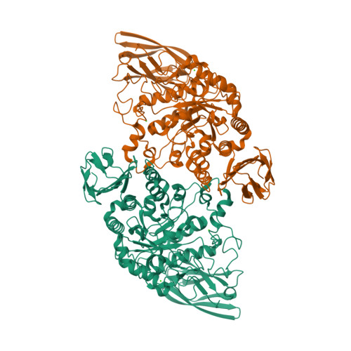
RCSB PDB - 3EDD: Structural base for cyclodextrin hydrolysis

3DNA Homepage -- Nucleic Acid Structures

RCSB PDB - 2C7O: HhaI DNA methyltransferase complex with 13mer  oligonucleotide containing 2-aminopurine adjacent to the target base  (PCGC:GMGC) and SAH
RCSB PDB - 2C7O: HhaI DNA methyltransferase complex with 13mer oligonucleotide containing 2-aminopurine adjacent to the target base (PCGC:GMGC) and SAH

RCSB PDB - 2N5O: Universal Base oligonucleotide structure

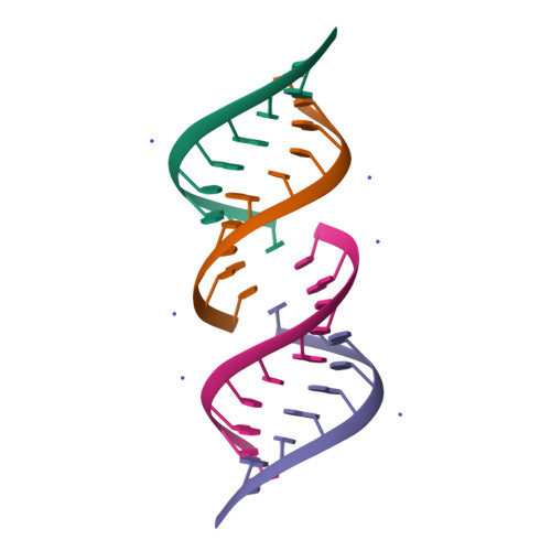
RCSB PDB - 5HNQ: Base Pairing and Structure Insights into the  5-Formylcytosine in RNA Duplex
RCSB PDB - 5HNQ: Base Pairing and Structure Insights into the 5-Formylcytosine in RNA Duplex

RCSB PDB - 1FUF: CRYSTAL STRUCTURE OF A 14BP RNA OLIGONUCLEOTIDE CONTAINING  DOUBLE UU BULGES: A NOVEL INTRAMOLECULAR U*(AU) BASE TRIPLE
RCSB PDB - 1FUF: CRYSTAL STRUCTURE OF A 14BP RNA OLIGONUCLEOTIDE CONTAINING DOUBLE UU BULGES: A NOVEL INTRAMOLECULAR U*(AU) BASE TRIPLE
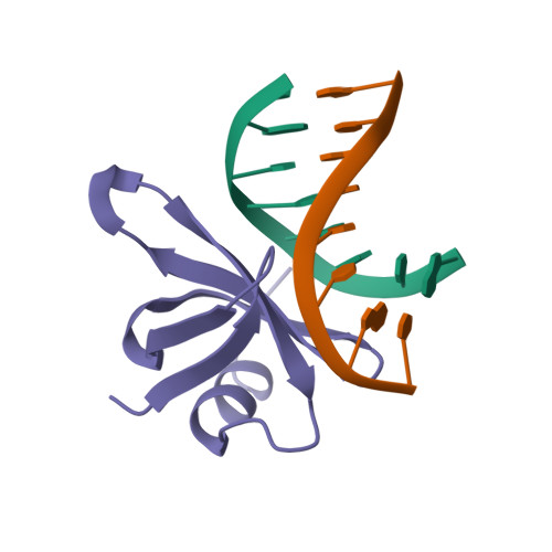
RCSB PDB - 1C8C: CRYSTAL STRUCTURES OF THE CHROMOSOMAL PROTEINS SSO7D/SAC7D  BOUND TO DNA CONTAINING T-G MISMATCHED BASE PAIRS
RCSB PDB - 1C8C: CRYSTAL STRUCTURES OF THE CHROMOSOMAL PROTEINS SSO7D/SAC7D BOUND TO DNA CONTAINING T-G MISMATCHED BASE PAIRS

RCSB PDB - 1QC0: CRYSTAL STRUCTURE OF A 19 BASE PAIR COPY CONTROL RELATED  RNA DUPLEX
RCSB PDB - 1QC0: CRYSTAL STRUCTURE OF A 19 BASE PAIR COPY CONTROL RELATED RNA DUPLEX

RCSB PDB - 1L3U: Crystal Structure of Bacillus DNA Polymerase I Fragment  product complex with 11 base pairs of duplex DNA following addition of a  dTTP and a dATP residue.
RCSB PDB - 1L3U: Crystal Structure of Bacillus DNA Polymerase I Fragment product complex with 11 base pairs of duplex DNA following addition of a dTTP and a dATP residue.
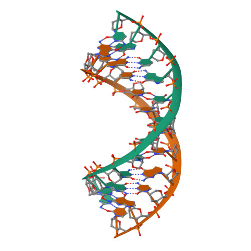
RCSB PDB - 1EC4: SOLUTION STRUCTURE OF A HEXITOL NUCLEIC ACID DUPLEX WITH  FOUR CONSECUTIVE T:T BASE PAIRS
RCSB PDB - 1EC4: SOLUTION STRUCTURE OF A HEXITOL NUCLEIC ACID DUPLEX WITH FOUR CONSECUTIVE T:T BASE PAIRS
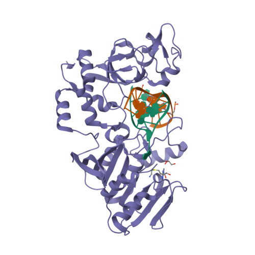
RCSB PDB - 7JY7: Structure of a 12 base pair RecA-D loop complex

RCSB PDB - 1R2L: A parallel stranded DNA duplex with an A-G mismatch base -pair

RCSB PDB - 1U48: Extension of a cytosine-8-oxoguanine base pair

RCSB PDB - 5HNQ: Base Pairing and Structure Insights into the  5-Formylcytosine in RNA Duplex
RCSB PDB - 5HNQ: Base Pairing and Structure Insights into the 5-Formylcytosine in RNA Duplex

RCSB PDB - 2IH2: Crystal structure of the adenine-specific DNA  methyltransferase M.TaqI complexed with the cofactor analog AETA and a 10  bp DNA containing 5-methylpyrimidin-2(1H)-one at the target base partner  position
RCSB PDB - 2IH2: Crystal structure of the adenine-specific DNA methyltransferase M.TaqI complexed with the cofactor analog AETA and a 10 bp DNA containing 5-methylpyrimidin-2(1H)-one at the target base partner position

RCSB PDB - 1DA9: ANTHRACYCLINE-DNA INTERACTIONS AT UNFAVOURABLE BASE BASE-PAIR  TRIPLET-BINDING SITES: STRUCTURES OF D(CGGCCG)/DAUNOMYCIN AND  D(TGGCCA)/ADRIAMYCIN COMPL
RCSB PDB - 1DA9: ANTHRACYCLINE-DNA INTERACTIONS AT UNFAVOURABLE BASE BASE-PAIR TRIPLET-BINDING SITES: STRUCTURES OF D(CGGCCG)/DAUNOMYCIN AND D(TGGCCA)/ADRIAMYCIN COMPL
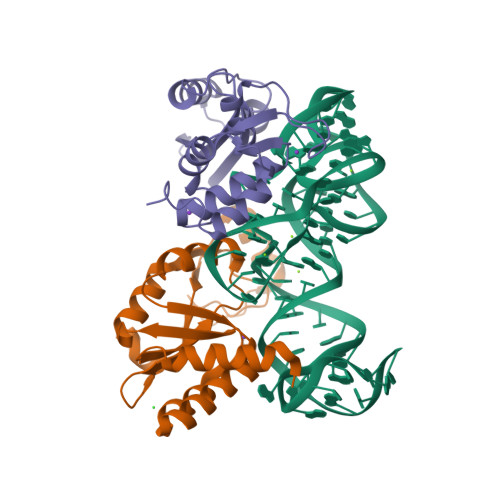
RCSB PDB - 1QCU: CRYSTAL STRUCTURE OF AN 18 BASE PAIR COPY CONTROL RELATED  RNA DUPLEX
RCSB PDB - 1QCU: CRYSTAL STRUCTURE OF AN 18 BASE PAIR COPY CONTROL RELATED RNA DUPLEX

RCSB PDB - 1U47: cytosine-8-Oxoguanine base pair at the polymerase active  site
RCSB PDB - 1U47: cytosine-8-Oxoguanine base pair at the polymerase active site

RCSB PDB - 5DAR: CRYSTAL STRUCTURE OF THE BASE OF THE RIBOSOMAL P STALK  FROM METHANOCOCCUS JANNASCHII
RCSB PDB - 5DAR: CRYSTAL STRUCTURE OF THE BASE OF THE RIBOSOMAL P STALK FROM METHANOCOCCUS JANNASCHII
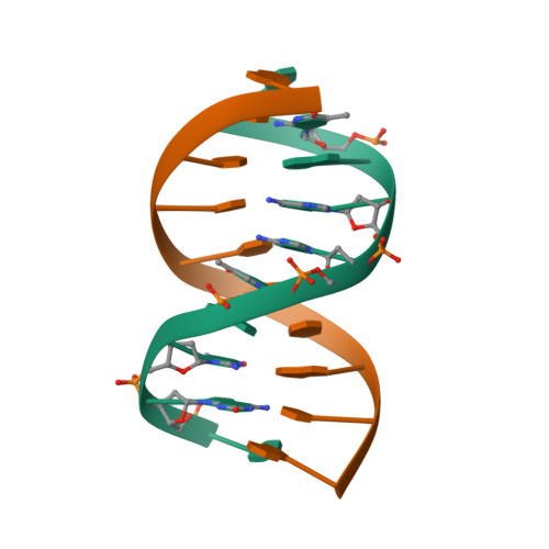
RCSB PDB - 1BE5: STRUCTURAL STUDIES OF A STABLE PARALLEL-STRANDED DNA  DUPLEX INCORPORATING ISOGUANINE:CYTOSINE AND ISOCYTOSINE:GUANINE BASE PAIRS  BY NMR, MINIMIZED AVERAGE STRUCTURE
RCSB PDB - 1BE5: STRUCTURAL STUDIES OF A STABLE PARALLEL-STRANDED DNA DUPLEX INCORPORATING ISOGUANINE:CYTOSINE AND ISOCYTOSINE:GUANINE BASE PAIRS BY NMR, MINIMIZED AVERAGE STRUCTURE

RCSB PDB - 1YFL: T4Dam in Complex with Sinefungin and 16-mer  Oligonucleotide Showing Semi-specific and Specific Contact and Flipped Base
RCSB PDB - 1YFL: T4Dam in Complex with Sinefungin and 16-mer Oligonucleotide Showing Semi-specific and Specific Contact and Flipped Base

RCSB PDB: Homepage
RCSB PDB: Homepage

RCSB PDB - 2BR0: DNA Adduct Bypass Polymerization by Sulfolobus  solfataricus Dpo4. Analysis and Crystal Structures of Multiple Base-Pair  Substitution and Frameshift Products with the Adduct 1,N2-Ethenoguanine
RCSB PDB - 2BR0: DNA Adduct Bypass Polymerization by Sulfolobus solfataricus Dpo4. Analysis and Crystal Structures of Multiple Base-Pair Substitution and Frameshift Products with the Adduct 1,N2-Ethenoguanine

RCSB PDB - 1P43: REVERSE PROTONATION IS THE KEY TO GENERAL ACID-BASE  CATALYSIS IN ENOLASE
RCSB PDB - 1P43: REVERSE PROTONATION IS THE KEY TO GENERAL ACID-BASE CATALYSIS IN ENOLASE